Facebookやってます

2011年12月17日土曜日

[アシュワース教授]パート5 圧−量ループ その3

その2の続き
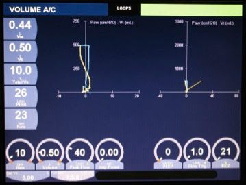
下の図のようなPVループの問題を修正する方法


Increase the Peak Inspiratory Flowrate
Many times an easy option to correct this type of patient-ventilator dys-synchrony is to increase the peak inspiratory flowrate so that the set flowrate now meets the patient’s demand. It is important to understand the ventilator to make sure that increasing the peak flowrate does not alter the tidal volume.


最高吸気流速を上げる
多くの場合、このタイプの患者−人工呼吸器非同調を修正するのに簡便な方法は最高吸気流速を上げて患者の需要に見合った吸気流速に設定することです。この場合、人工呼吸器の特性を理解し、吸気流速設定を上げることで換気量が変化することがないように確認することが重要です。


Change to a Rectangular Flow Waveform
When ventilating a patient in volume-targeted ventilation, it is possible on most ventilators to select a decelerating or a rectangular flow waveform. If a decelerating flow waveform is selected, the flowrate gradually decreases throughout inspiration. In some patients, this reduction in flowrate may decrease below the patient’s actual inspiratory flowrate, resulting in patient-ventilator dys-synchrony and the Figure on the PV Loop. If the flow waveform is changed to a rectangular flow waveform, the ventilator will deliver the set peak flowrate throughout inspiration, possibly reducing the patient-ventilator dys-synchrony.


流速波形を矩形波へ変更する
量規定換気の場合、ほとんどの人工呼吸器では漸減波と矩形波のどちらかを選択することができます。漸減波を選択すると、吸気相全般に渡って吸気流速が低下するため、患者によっては実際の吸気流速を下回るところまで減少し、図のような患者−人工呼吸器の不同調に至ることがあります。矩形波へ変更すると、人工呼吸器は吸気全般に渡って設定された一定の吸気流速を維持するため、患者−人工呼吸器非同調を緩和する可能性があります。


Increase the set Tidal Volume
Increasing the set tidal volume may correct the situation. However, it is very important to make sure that the tidal volume is not too high as it may increase the chance of ventilator-induced lung injury.


設定1回換気量を増加させる
設定1回換気量を増加させることで解決できることがあります。しかし、1回換気量を高くしすぎることで、人工呼吸器による肺損傷(ventilator-induced lung injury:VILI)の危険性を高めることのないよう確認することが重要です。


Change to a different Ventilator Mode
Another option is to change the mode of ventilation. If the patient is changed to a pressure-targeted mode of ventilation, the patient will now have control over the inspiratory flowrate. This generally results in an improved patient-ventilator synchrony.


換気モードを変更する
もう一つの方法は、換気モードを変更することです。圧規定換気に変更すれば、患者は吸気流速の規定を受けず、自分でコントロールすることができます。通常は圧規定換気に変更することで患者−人工呼吸器同調性は改善します。


Use of Medications to Improve Patient-Ventilator Synchrony
Medications can be used to improve patient-ventilator synchrony. However, it is important that the patient is not just automatically heavily sedated. Increasing emphasis is being placed on not sedating the patient as much as was done in the past. Instead, appropriate use of pain control and control of anxiety are encouraged. 


患者−人工呼吸器同調性を高めるために薬剤を使用する
患者−人工呼吸器同調性を高めるために薬剤を用いることも可能です。しかし、安易に患者を深い鎮静下に置かないことが重要です。過去に我々がしてきたような過鎮静をしないことが、ますます強調されてきています。その代り、適切な鎮痛と不安への対処が奨励されます。


The next post will discuss “beaking” in a PV Loop that may be caused by over-distension or by a patient activating his expiratory muscles while the ventilator is still delivering a breath.

次回の投稿ではPVループにおける“ビーキング”(くちばし様波形)について述べます。これは過膨張もしくは人工呼吸器がまだ吸気を行っている間に、患者が呼気を開始しようと呼気筋を活動させた場合にみられるものです。 


日本語訳 岩本志津(米国呼吸療法士)

2011年12月16日金曜日

[アシュワース教授]パート5 圧−量ループ その2

お待たせしました。Lonny Ashworth教授のグラフィックレクチャー パート5-2です。前回に引き続き、圧ー量ループの話です。今回から異常波形の説明になります。

ーーーーーーーーーーーーーーーーーーーーーーーーーーーーーーーーーーーーーーーーーーー
As discussed in the previous post, “Part 5 – Pressure-Volume Loop”, the Pressure-Volume Loop (PV Loop) can be used to detect patient-ventilator dys-synchrony. It is important to monitor patients closely to detect patient-ventilator dys-synchrony because this may lead to ventilator-induced lung injury, increased metabolic rate and may even prolong the number of days the patient receives mechanical ventilation.

前回の「パート5 圧‐量ループ」にあるように、圧−量ループ(PVループ)は患者−人工呼吸器非同調の発見に用いることができます。患者−人工呼吸器非同調を密に観察することは重要です。なぜなら肺損傷、代謝率の増加、ひいては人工呼吸器装着期間の延長につながる可能性があるからです。


When the patient’s inspiratory flowrate exceeds the set flowrate during volume targeted ventilation, the airway pressure does not increase as expected, and it actually may decrease. This was discussed in an earlier post discussing the Pressure versus Time waveform, and the waveform is referred to as a Sagging Pressure Versus Time Waveform.

量規定換気において、患者の吸気流速が設定吸気流速を上回った場合、気道内圧は予想される程度までは上昇せず、実際低下することもあります。この現象は過去の圧−時間波形の投稿でも述べましたが、陥没(Sagging)圧−時間波形といいます。


If you observe the PV Loop when a patient’s inspiratory flowrate exceeds the set flowrate, you will notice that the pressure begins to decrease during inspiration and then generally increase at the end of inspiration when the patient exhales. This results in a tracing that is similar to the Figure.

臨床現場でこのようなPVループを観察すると、吸気時に圧がまず低下し、次に吸気の終わりから呼気に移行するときに上昇することに気付くでしょう。この場合、図のような波形となります。


When you look at the tracing below, you can see how the pressure gradually decreased as the volume was being delivered, then increased at the end of inspiration, resulting in the Figure.

図の波形では、規定した換気量が供給される間、圧が徐々に低下し、吸気終末で圧が上昇していることが見て取れます。

When you observe the Figure on a PV Loop, you must try to determine why the patient’s flowrate exceeds the set flowrate. There are many options to correct this situation, however, it is important for the clinician to determine which option is best for that specific patient at that specific point in time.

のようなPVループを見た場合、患者の吸気流速が設定吸気流速を上回る理由を判断する必要があります。この状況を修正する方法はいくつもありますが、それぞれのタイミングでそれぞれの患者に適した方法を選ぶことが重要です。


日本語訳 岩本志津(米国呼吸療法士)


2011年11月23日水曜日

第4回ワークショップ終了

 2011年11月19、20日に第4回若手医師のための人工呼吸器ワークショップが行われました。今回も熱心な方々に受講していただき、充実した2日間となりました。受講された方からコメントの一部を紹介します。
  • 自分で実際にモードの違いを体験してみることは重要で役立ちました。
  • 自分の人工呼吸管理がどれだけ患者に苦痛を与えていたかがわかりました。
  • 難しかったですが、レスピレーターに触って、いろいろな状況を考えながらできてよかったです。
  • ロールプレイはやはりとてもためになる。
  • 実戦向きでとてもわかりやすかったです。
  • とても勉強になる2日間であり、楽しく学ぶことができました。明日からでも自分の病院で積極的に呼吸器をいじって患者さんに良い呼吸を提供したいと思います。

 みなさん、大変お疲れ様でした。ご自身の施設に戻られた後も、どんどん仲間を増やして、より良い呼吸ケアを提供して下さい。

人工呼吸器実習

人工呼吸器実習

人工呼吸器実習

人工呼吸器実習

NPPV実習

NPPV実習

今回のスタッフ

 次回以降のワークショップの予定は決まり次第お知らせいたします。


2011年10月16日日曜日

沖縄呼吸ケア研究会のご紹介

2005年3月、沖縄県内の呼吸療法認定士が発足させ、急性期から慢性期・在宅を含めた呼吸ケアのレベル向上を目的としている研究会です。
年に2回のセミナーや、月に1回の勉強会を開催しています。
先日沖縄に行ったとき、研究会の集まりに参加させていただきました。みなさん、とても楽しい方々で、呼吸ケアだけでなくいろんな話で盛り上がっていました。
彼らはホームページももっていますので、是非立ち寄ってみてください。
研究会の皆様、今後ともよろしくお願いします!

2011年9月30日金曜日

ワークショップに参加された皆様へ

 第1〜3回若手医師のための人工呼吸器ワークショップ、人工呼吸器ブラッシュアップワークショップにご参加下さった皆様。

 すでにメールでご案内しておりますが、今後のワークショップの質をさらに改善するために、これまで受講された方を対象に、アンケートをさせて頂いております。
 ワークショップ受講前後で人工呼吸器の使用方法に変化があったのか、どのような講義が役に立ったのかをお聞きしています。約1分でできる内容ですので、まだご回答頂いていない方がいらっしゃいましたら是非ご協力をお願いします。
 皆さんの貴重なご意見を参考に、ワークショップをさらに実りのあるものにしたいと考えております。お忙しい中、お願いして申し訳ありませんが、よろしくお願いします。
 

第1回若手医師のための人工呼吸器ワークショップ
(亀田総合病院シミュレーションセンターにて)

第2回若手医師のための人工呼吸器ワークショップ
(コヴィディエンジャパン株式会社 板橋事務所にて)

第3回若手医師のための人工呼吸器ワークショップ
(コヴィディエンジャパン株式会社 板橋事務所にて)

人工呼吸器ブラッシュアップワークショップ
(コヴィディエンジャパン株式会社 板橋事務所にて)

2011年9月19日月曜日

[アシュワース教授]パート5 圧−量ループ


Part 5 – Pressure-Volume Loop
パート5 圧−量ループ

The Pressure-Volume Loop is used to help identify patient-ventilator dys-synchrony and over-distension, which may be caused by using too large of tidal volume or excessive PEEP. Some published research has identified patient-ventilator dys-synchrony in up to 25% of patients receiving ventilatory support. Patient-ventilator dys-synchrony may increase the patient’s work-of-breathing, prolong ventilatory support and may increase the chance of ventilator-induced lung injury. Therefore, it is very important to carefully monitor the Pressure-Volume Loop to prevent patient-ventilator dys-synchrony.

圧−量ループは、患者−人工呼吸器非同調や、大きすぎる一回換気量または高すぎるPEEPによって引き起こされうる過膨張を発見するのに使います。いくつかの研究では、人工呼吸管理をされている患者の25%までに患者−人工呼吸器非同調が見られたとしています。患者−人工呼吸器非同調があると、患者の呼吸仕事量が増加し、人工呼吸器が必要な期間を長引かせる可能性があり、人工呼吸器誘発の肺障害(VILI)のリスクを増大させる可能性があります。よって患者−人工呼吸器非同調を防ぐために、圧−量ループを注意深くモニターすることが大変重要になります。

Normal Pressure-Volume Loop
正常な圧−量ループ

The following images are examples of a patient being ventilated in Volume-Targeted ventilation. The ventilator settings are: Volume A/C; Respiratory Rate 20/minute; Tidal Volume 0.5 L; Peak Flowrate 60 LPM; Square Flow Waveform; PEEP 0 cm H2O. This patient is not making any spontaneous efforts.

次に示す画像は量規定換気で換気されている患者の例です。人工呼吸器の設定は量規定アシストコントロール、呼吸回数20回/分、一回換気量0.5L、最高気道流速60LPM、矩形波、PEEP 0 cm H2Oです。この患者は自発呼吸の努力を全くしていません。

Pressure is being graphed on the X-axis; Volume is being graphed on the Y-axis. At the beginning of inspiration, pressure starts at 0 cm H2O because PEEP is set at 0 cm H2O. As the 500 ml tidal volume is delivered, the airway pressure increases to approximately 19 cm H2O. The inspiratory tracing is red because the patient did not initiate the breath. Expiration is graphed as blue-green on this ventilator.

圧はX軸に示されており、量はY軸に示されています。PEEP0 cm H2Oに設定されているために、吸気の始まりでは圧は0 cm H2Oになっています。500mlの一回換気量が送られたとき、気道内圧は約19 cm H2Oまで上昇しています。患者が吸気を開始していないために、吸気の軌跡は赤で示されています。この人工呼吸器上では呼気は青で示されています。

自発呼吸がないときの圧−量ループ

In the following graph, directional arrows were added to the image to demonstrate that inspiration is graphed going upward and to the right; during expiration, the tracing goes toward the left and downward.

次に示すグラフでは、吸気時に軌跡が右上方に上がって行き、呼気時に左下方へ下がっていくのを示すために、矢印を加えています。

吸気(Inspiration)、呼気(Expiration)を矢印で示しています

In this graph, lines were added to demonstrate the peak inspiratory pressure of approximately 19 cm H2O and the tidal volume of 500 ml.

このグラフでは最高吸気圧が約19 cm H2Oであり、一回換気量が500 mlであることを示すために線を加えています。

最高吸気圧19cmH2O、1回換気量500mlであることを示しています

When you click on the attached video, it will play a video file of a normal Pressure-Volume Loop. Please note that the inspiratory tracing is yellow, because the patient is triggering assisted breaths.

下に添付したのは正常な圧−量ループのビデオです。患者が補助呼吸をトリガーしているために吸気の軌跡が黄色であることに注目してください。


The next two posts about graphics will discuss abnormal Pressure-Volume Loops.

次の2回分のグラフィック講義では、圧−量ループの異常についてお話します。

日本語訳 マクマーン由香(米国呼吸療法士)

2011年9月18日日曜日

[アシュワース教授]次回予告

 Lonny Ashworth教授による次回のグラフィックレクチャーでは、P-Vループを扱います。7月の「グラフィックセミナーの夕べ」でも扱いましたが、ループは直感的にわかりにくいと、苦手意識を感じておられる方もいらっしゃるようです。
 いつものように、非常にわかりやすい説明をしてくださると思いますので、どうぞお楽しみに。


2011年9月14日水曜日

20秒でできる人工呼吸器アンケート第3弾 集計

 今回も多数の皆様から回答を頂きました。
 ルーチンで使っていらっしゃる方は少ないのですね。適応に応じて使っていらっしゃるのはすばらしいです。うちの施設では、いまだに何となくオーダーされているネブライザー指示が多く、「ルーチンではないが、投与することが多い」に当てはまると思います。大いに改善の余地ありです。
 気管支拡張剤以外に投与する薬剤としては、ビソルボンやムコフィリンと言った去痰薬の使用が多く、その他にはネオフィリン、インタールを使うという方もいらっしゃいました。どれくらい「効いている」感があるのか、使用している方の印象も是非お聞きしたいです。
 ラシックス、モルヒネ、ヘパリンなどの吸入を使っておられる方がいるのではないかと期待していましたが、回答はありませんでした。
 皆様、ご協力ありがとうございました。

2011年9月6日火曜日

20秒でできる人工呼吸器アンケート第3弾

 「人工呼吸管理中の患者さんに、ネブライザーってやらないとだめなの?」という質問を頂くことがあります。手間的にも感染管理的にも、やらないで済むならその方が良さそうですよね。
 と言うわけで、20秒でできる人工呼吸器アンケート第3弾はネブライザーについてです。是非ご参加下さい。


2011年9月3日土曜日

[アシュワース教授] パート4 量-時間波形


Part 4 – Volume versus Time Waveform
パート4 量-時間波形

The Volume versus Time Waveform is used to identify the presence of a leak in the system. This leak may be caused by a leak inside the ventilator, a leak in the circuit, a leak within the endotracheal tube or tracheostomy tube cuff, or even a pleural leak. Even though the Volume versus Time Waveform can identify the presence of a leak, it does not identify where the leak is located; it is up to the clinician to determine the source of the leak.

 量-時間波形はシステム内のリークを認識するために用いる。リークの発生する箇所には、人工呼吸器の本体内部、回路、挿管チューブや気管切開チューブのカフ、胸腔ドレーンなどがある。量-時間波形でリークの存在が確認できても、リークが発生している箇所を特定することはできないため、臨床家がリークの箇所を突き止めなければならない。


The ventilator has two flow sensors. One flow sensor is located within the ventilator; it measures the inspiratory flow and calculates the inspiratory volume. The second flow sensor is located after the exhalation valve; it measures the expiratory flow and calculates the expiratory volume. When a leak occurs, the inspiratory volume is not affect, however, because of the leak, some of the gas does not pass through the expiratory flow sensor, resulting in a change in the Volume versus Time Waveform.

 人工呼吸器には2つのフローセンサーがある。ひとつは人工呼吸器の内部にあり、吸気フローを計測して吸気換気量を計算する。もうひとつのフローセンサーは呼気バルブの後に位置しており、呼気フローを計測して呼気換気量を計算する。リークが発生すると、呼気換気量は影響をうけないが、ガスの一部は呼気フローセンサーを通過しないため、量-時間波形に変化が起こることになる。


The following image is an example of a patient being ventilated in Volume-Targeted ventilation. The ventilator settings are: Volume A/C; Respiratory Rate 20/minute; Tidal Volume 0.5 L; Peak Flowrate 60 LPM; Square Flow Waveform; PEEP 0 cm H2O. This patient is not making any spontaneous efforts. The Volume versus Time Waveform is the bottom tracing. It gradually increases to the set tidal volume of 0.5 L, then returns to baseline at the end of expiration. This is a normally appearing Volume versus Time Waveform.

 次に表示する画像は、量規定換気で管理されている患者である。人工呼吸器設定は、量規定アシストコントロール、呼吸回数20/分、一回換気量500ml、ピークフロー 60LPM矩形波、PEEP 0cmH2Oである。患者の自発呼吸はない。3つの波形のうち最下段が量-時間波形である。波形は500mlの換気量に達するまでなだらかに上昇した後、呼気へ移行し呼気終末で基線に戻る。これが正常の量-時間波形である。 

正常の量-時間波形


When you look at the Volume versus Time Waveform in the following tracing, you notice that the waveform appears normal in the first two breaths. The inspiratory volume and expiratory volume are both approximately 500 mL. However, when you look at the third and fourth breaths, you notice that the inspiratory volume is 500 mL, but the expiratory volume does not return all the way to baseline. In this case, there is a leak of approximately 150 mL. This can also be confirmed by looking at the digital display of the exhaled tidal volume, which shows 0.35 L or 350 mL. Because the set tidal volume is 0.5 L, the leak is approximately 150 mL.

 次の量-時間波形を見てほしい。最初の2呼吸は正常波形であり、吸気、呼気の換気量ともに500mlであることを示している。しかし後半2つの波形では吸気換気量は500mlだが、呼気換気量は完全に基線に戻っておらず、およそ150mlのリークがあることがグラフィックから確認できる。このことは、画面左端の呼気換気量(Vte)のデジタル表示に0.35L(すなわち350mlと表示されていることからも確認できる。設定換気量は500mlなので、リーク量はおおよそ150mlとなる。

後半の2つの波形ではリークが見られる


When a leak is detected, it is important that the clinician identifies the leak and corrects the leak, if possible. If the leak is inside the ventilator, within the circuit or due to a cuff leak, the patient will not receive the set tidal volume and the alveolar ventilation will be reduced. Many times the leak can be identified by listening closely around the circuit or holding your hand near the circuit. If the leak is in the cuff, frequently an audible sound can be heard as gas passes around the cuff during inspiration. Sometimes it may be necessary to place your stethoscope on the patient’s trachea to identify a cuff leak. If the leak is due to a pleural leak and the patient has a properly functioning chest drainage system, it is important to make sure the amount of leak is monitored closely.

 リークが見つかった場合、どこでリークが発生しているのかを判断し、可能なら対処することが重要である。人工呼吸器内部、回路、カフにリークがあると、患者は設定された換気量を得ることができず、肺胞換気量が減少する。回路にリークがある場合、回路の近くで耳を澄ます、もしくは回路に手をかざすことでリークの部位を見つけられることが多い。もしカフ周囲でリークが存在するのであれば、吸気時にガスがカフ周囲を通る音で判断できる。時には、聴診器を患者の気管にあてて聴いて、カフリークを判断するのが必要なこともある。胸腔ドレーンからのリークがある場合には、実際のリーク量を密にモニターすることが重要である。

日本語訳 岩本志津(米国呼吸療法士)

2011年8月28日日曜日

20秒でできる人工呼吸器アンケート第2弾 集計

 今回も多数のご回答ありがとうございます。人工呼吸器離脱に関するアンケートの集計です。
 ゆっくり下げる派とSBT派がちょうど同数くらいですね。
 ゆっくり下げる派ではCPAP+PSが最多で、次いでSIMVとなっています。
 SBTでもCPAP+PSが最多となっています。皆さん、どれくらいのPSをかけているのでしょうか?


2011年8月24日水曜日

20秒でできる人工呼吸器アンケート第2弾です

 皆さんは普段どのように人工呼吸器からの離脱を行っていますか?SIMVでじっくり人工呼吸器設定を下げていく派ですか?それとも、とりあえずSBTをやってみる派でしょうか?
 人工呼吸器離脱の方法について簡単なアンケートを行いたいと思いますので、ご協力よろしくお願いします。

2011年8月20日土曜日

ワークショップの申し込みを締め切りました

 第4回若手医師のための人工呼吸器ワークショップの申し込みを締め切りました。


 皆様からは、申し込みと共に「こんなことを学びたい!」という高い目的意識の籠もったコメントを多数頂きました。若手医師の先生方が、呼吸ケアにこれほど興味を持っておられることを大変うれしく思います。できれば申し込み下さった方全員に参加して頂きたいのですが、今回は申し込み多数のため抽選となります。抽選の結果はメールにてお知らせいたします。


 今回参加することになる方々とは、ワークショップで熱く呼吸器話をすることを楽しみにしております。残念ながら今回抽選からもれた方とは、次回以降のワークショップで是非お会いしたいと思います。

2011年8月12日金曜日

[アシュワース教授] パート3その3 見逃された吸気努力を見つけるためのフロー−時間波形

Lonny Ashworth教授によるグラフィック講義のパート3その3です。
ーーーーーーーーーーーーーーーーーーーーーーーーーーー


The Flow versus Time Waveform can be used to detect missed efforts, frequently called missed breaths or non-triggered efforts or non-triggered breaths. This means that the patient creates and inspiratory effort but the effort is not sufficient enough to trigger a breath from the ventilator.  It is also possible, and many times easier, to identify missed breaths by comparing the patient’s actual respiratory rate with the ventilators rate. If you count the patient’s respiratory rate and it is greater than the respiratory rate delivered by the ventilator, the patient has missed efforts.

 フロー対時間波形は、見逃された吸気努力を見つけるために使用します。それはしばしば見逃された呼吸、もしくはトリガーされない吸気努力またはトリガーがされない呼吸とも呼ばれます。つまり患者は吸気努力をしますが、その努力は人工呼吸器をトリガーするのには十分でないという事になります。またもっと簡単に、患者の実際の呼吸回数と人工呼吸器の設定呼吸回数を比較することによって、見逃された呼吸を見つけ出す事も可能です。もしあなたが呼吸回数を数えて、それが人工呼吸器により送られる呼吸回数よりも多いならば、 見逃された吸気努力があることになります。


There are three primary causes of missed efforts. One likely cause of missed efforts is a poorly set sensitivity. The pressure and flow sensitivity should always be set as sensitive as possible, without allowing the ventilator to auto-trigger, thereby delivering an assisted breath when the patient did not create an inspiratory effort. Generally, the pressure sensitivity should be set at 1 cm H2O and the flow sensitivity should be set at around 2 or 3 LPM.

 見逃された吸気努力には主な原因が3つあります。 一つ目は不適当な感度設定です。圧もしくはフロー感度はできるだけ鋭敏に設定しますが、鋭敏にしすぎてオートトリガーが起こり、患者が吸気努力をしていない時にも補助呼吸が送られるということがないようにします 。一般的に圧感度は1 cm H2O、フロー感度は23 L/程度に設定します。


The second major cause of missed efforts is AutoPEEP. AutoPEEP was discussed previously and is caused by an increased airway resistance or decreased expiratory time. When patients have AutoPEEP, the increased airway resistance prevents the patient from exhaling their air to their baseline functional residual capacity. If possible, reduce the AutoPEEP by decreasing the airway resistance or increasing the expiratory time. Another option is too very slowly increase the set PEEP by 1 cm H2O at a time, watching the trigger delay and the number of missed breaths. When the set PEEP is close to the AutoPEEP, the trigger delay (the amount of time from when the patient begins the inspiratory effort to when the ventilator actually delivers a breath) and missed breaths should be reduced.

 見逃された吸気努力の主な原因の二つ目は、AutoPEEP(オートピープ)です。 以前にも述べましたが、AutoPEEPは気道抵抗の上昇もしくは呼気時間の短縮によって起こります。AutoPEEPが存在する時、上昇した気道抵抗のため患者はFRC(機能的残気量)まで息を吐くことができません。もし可能であれば気道抵抗を下げるか、呼気時間を延長する事によりAutoPEEPを下げます。その他に、トリガー遅延(患者が吸気努力を始めてから、人工呼吸が実際に吸気を供給するまでの時間)と見逃された呼吸数を見ながら、PEEP1 cm H2Oずつゆっくりと上げていく方法があります。設定PEEPAutoPEEPに近づくと、トリガー遅延と見逃された呼吸は減少します。


The third major cause of missed efforts is due to weakened or fatigued inspiratory muscles and a weak drive to breathe. If the patient’s inspiratory muscles are so weak or drive to breathe is so suppressed, the patient may not be able to generate sufficient effort to trigger a breath. If this appears to be the case, check the trigger sensitivity to be sure that it is set as sensitive as possible.

 見逃された呼吸の主な原因の三つ目は、吸気筋力の低下または疲労と、呼吸ドライブの低下です。患者の吸気筋力が弱いか、呼吸中枢が抑制されている場合、患者は吸気をトリガーするに十分な呼吸努力を起こす事ができないことがあります。この場合は トリガー感度をチェックし、可能な限り感度を鋭敏に設定します。


The following image is an example of a patient being ventilated in Volume-Targeted ventilation. The ventilator settings are: Volume A/C; Respiratory Rate 20/minute; Tidal Volume 0.5 L; Peak Flowrate 60 LPM; Decelerating Flow Waveform; PEEP 0 cm H2O.
The Flow versus Time Waveform is the second tracing. Notice the yellow elliptical circles marking the missed efforts. The patient tried to initiate a breath, was unsuccessful and exhaled. So, when you look at the Flow versus Time Waveform and see additional expiratory deflects on the expiratory flow waveform, caused by additional expiratory efforts without any inspiratory flow from the ventilator, the patient has experienced a missed effort.

 次に示す画像は、量規定換気により換気されている例を示しています。人工呼吸器の設定は量規定アシストコントロール、呼吸数20回/分、一回換気量0.5L、最高流量60L/分、漸減波、PEEP 0cmH2Oです。
 上から2番目がフロー対時間波形で、黄色の楕円で囲まれているのが見逃された努力です。患者は吸気を開始しようとしていますが、不成功に終わっており、呼気が起こっています。人工呼吸器からの送気がないのに、フロー対時間波形において呼気努力による呼気波形の揺れがある場合には、見逃された呼吸努力があります。


日本語訳 マクマーン由香(米国呼吸療法士)

2011年8月11日木曜日

人工呼吸器アンケート結果

 先日行った「20秒でできる人工呼吸器アンケート」の結果です。119人の方にお答え頂きました。


 やはりSIMVが多いですね。SIMV、A/CともにPCVで使う人の方がVCVより多いのはちょっと驚きでした。みなさんの印象はいかがでしょうか?

2011年8月8日月曜日

20秒でできる人工呼吸器アンケート

みなさんの人工呼吸器使用状況をお聞きしたいと思います。簡単なアンケートですのでご協力よろしくお願いします。

2011年8月3日水曜日

若手医師のための人工呼吸器ワークショップ(ブラッシュアップ含む)中の風景


自己紹介から始まったワークショップ。皆様とっても学習意欲があり、質問も積極的で、私自身もその質問から学ばせていただきました。このような学習の機会は、伝える側も受ける側もそれぞれに得ることがあり、生涯学習者である医療従事者にとっては無くてはならないものだと思います。ご参加くださったみなさま、ありがとうございました。仕事に戻って、患者への治療、後輩への指導、コメディカルとの関わりなどで、すこしでも「そういえばこういうことやってたな」みたいに思い出していただけると幸いです。

2011年7月27日水曜日

メーリングリスト「呼吸ケア倶楽部」開設のお知らせ

 かねてより希望の多かった呼吸ケアに関するメーリングリストを開設いたします。
 医師だけではなく、呼吸ケアに携わるすべての職種の方に参加頂きたいと考えております。日常診療における疑問や、興味深い症例、各施設での取り組みなど様々な情報を共有していきたいと思いますので、是非お誘い合わせの上ご加入下さい。


 参加を希望される方はこちらからどうぞ。


 また写真、ビデオなどを共有するためFacebookページ「呼吸ケア倶楽部」も開設いたしました。こちらも併せてご利用下さい。

2011年7月16日土曜日

人工呼吸器グラフィックの夕べ 申し込みを終了しました

 先日案内しました「人工呼吸器グラフィックの夕べ」ですが、おかげさまで多数のお申し込みを頂きましたので、受付を終了いたします。ありがとうございました。
 お申し込み下さった皆様とは当日お会いできることを大変楽しみにしております。
 夕食は出ませんので、各自持ち込みでお願いします。

2011年6月26日日曜日

看護師以外にも役に立つ内容掲載 第2弾


前回の看護雑誌「重症集中ケア」の特集、エキスパートの呼吸器ケアの続編が先日発売になりました。この雑誌は、年間購読ですので書店でお買い求めすることはできませんが、所属施設の看護師で雑誌をお持ちの方がいらっしゃったら、是非内容を共有していただければと思います。

今回号には、当ワークショップに携わっていただいている方々も執筆しています。


人工呼吸管理中の吸入
戎 初代 杏林大学医学部付属病院 高度救命救急センター 集中ケア認定看護師

加温加湿
石井宣大 東京慈恵会医科大学附属第三病院 臨床工学部 係長

体位管理
小松由佳 東京慈恵会医科大学附属病院 看護師長/集中ケア認定看護師

気管支鏡検査時の準備と介助
戎 初代 杏林大学医学部付属病院 高度救命救急センター 集中ケア認定看護師

ARDS患者の管理
田中竜馬 LDS Hospital Pulmonary and Critical Care Medicine

COPD患者の管理
長谷川景子 医療法人鉄蕉会 亀田総合病院 呼吸器内科 スタッフ ほか

酸素療法
露木菜緒 杏林大学医学部付属病院 集中ケア認定看護師

NPPVの基礎知識
小原史子 フィリップス・レスピロニクス合同会社 マーケティング部
スーパーバイザー/看護師/米国呼吸療法士


NPPV使用の実際
濱本実也 公立陶生病院 ICU 看護師長/集中ケア認定看護師

早期離床
齋藤 洋 医療法人鉄蕉会 亀田総合病院 リハビリテーション室 理学療法士 ほか

初期の情報収集
マクマーン由香 医療法人社団幸徳会 薬袋内科クリニック
通所リハビリテーションセンター エム センター長


2011年5月17日火曜日

[アシュワース教授] パート3その2 オートPEEPを発見するためのフロー−時間波形

前回の続きです

When you look at the Flow versus Time Waveform in the following image, you will notice that the expiratory flow does not return to baseline before the next breath begins. Therefore, the patient has AutoPEEP.

 以下の図中のフロー−時間波形を見た時、呼気のフローが次の吸気が始まる前に基線に戻っていないのに気づくでしょう。つまりこの患者にはオートPEEPが起こっています。


Sometime it is difficult to determine whether or not AutoPEEP is present and it is helpful to change the scale of the Flow versus Time Waveform. In the following image, the scale was changed to allow the clinician to more closely examine the Flow versus Time Waveform, and determine that AutoPEEP is present, as expiratory flow does not return to baseline before the next breath begins.

 時々、オートPEEPが存在するか否か判断が困難な時があります。その時はフロー−時間波形のスケールを変えるとよいでしょう。次の図の中では、呼気フローが次の吸気が始まる前に基線に戻っていない時、臨床家がフロー−時間波形をより詳細に観察でき、オートPEEPが存在するかどうか調べられるようにスケールが変えています。


It is not possible to measure the AutoPEEP with the Flow versus Time Waveform. You can only determine whether or not AutoPEEP is present. If the clinician wants to measure the AutoPEEP, the clinician can provide an expiratory pause (expiratory hold) and measure the airway pressure at the beginning of the next breath. During this procedure, the patient must not make any inspiratory efforts.

 フロー−時間波形でオートPEEPの値を測定することはできません。オートPEEPが存在するかどうかを判断するだけです。もし、臨床家がオートPEEPの値を測定したいならば、呼気ポーズ(呼気ホールド)を使用し、次の吸気の始めの気道内圧を測定することができます。この過程において、患者は吸気努力をしてはいけません。

As mentioned previously, AutoPEEP may be reduced by decreasing airway resistance or increasing expiratory time. In most modes it is possible to increase the expiratory time by decreasing inspiratory time or decreasing the respiratory rate.

 上で述べたように、気道抵抗を低下させること又は、呼気時間を延長させることでオートPEEPを減少させることができます。ほとんどのモードで吸気時間を減少させる又は呼吸回数を減少させることで呼気時間を延長させることが可能です。


日本語訳 マクマーン由香(米国呼吸療法士)

2011年5月8日日曜日

[アシュワース教授] パート3その1 オートPEEPを発見するためのフロー−時間波形


The Flow versus Time Waveform can be used to detect the presence of AutoPEEP. AutoPEEP, frequently called air-trapping, results when the patient is not given enough time to exhale back to baseline. This results in an increased Functional Residual Capacity (FRC), increased mean airway pressure and may make it difficult for the patient to trigger additional breaths from the ventilator.

 フロー時間波形はオートPEEPが存在するかを見る時に使用することができます。オートPEEPはしばしばエアートラッピングと呼ばれますが、基線に戻るまでの十分な呼気時間が患者に与えられない時に起こります。これはFRC(機能的残気量)の増加を引き起こし、平均気道内圧を上昇させます。そして患者が人工呼吸器からの呼吸をトリガーすることを困難にさせてしまう可能性があります。


When patients have an increased airway resistance (possibly due to bronchospasm, secretions, small endotracheal tube, mucosal edema) or an increased compliance (possibly due to emphysema), they are more likely to have AutoPEEP, if the expiratory time is not long enough. Methods of reducing AutoPEEP include reducing airway resistance and lengthening the expiratory time. In most ventilator modes, the expiratory time can be increased by decreasing the inspiratory time or decreasing the respiratory rate.

 患者の気道抵抗が増加した時(気管支攣縮、気管支内分泌物、小さい気管内チューブ、粘膜浮腫等のため)、またはコンプライアンスが増加した時(肺気腫等のために)、呼気時間が十分長くない場合はオートPEEPを起こしやすくなります。オートPEEPを減少させる方法としては気道抵抗を減少させる事と呼気時間を長くする事が挙げられます。ほとんどの人工呼吸器のモードでは吸気時間を減少させるか、または呼吸回数を減少させることで呼気時間を長くすることができます。


The following image is an example of a patient being ventilated in Volume-Targeted ventilation. The ventilator settings are: Volume A/C; Respiratory Rate 20/minute; Tidal Volume 0.5 L; Peak Flowrate 60 LPM; Decelerating Flow Waveform; PEEP 0 cm H2O. This patient is not making any spontaneous efforts.

 以下の図は容量規定換気によって換気されている患者の例です。人工呼吸器の設定は、容量規定のアシストコントロール、呼吸回数20回/分、一回換気量0.5L、最高流速60LPM、漸減波、PEEP 0 cm H2Oです。この患者は自発呼吸の努力をしていません。


The Flow versus Time Waveform is the second tracing. Notice how the tracing on the graph goes up to the set peak flowrate of 60 LPM, then gradually decreases throughout inspiration because the decelerating ramp flow waveform was selected. When expiration begins, the tracing demonstrates the peak expiratory flow of approximately 50 LPM, and then the tracing gradually returns back up to baseline at the end of expiration. Because the expiratory flow returns to baseline, the patient has no AutoPEEP.

 上から2つめがフロー−時間波形です。漸減波形が選択されているためにグラフ上のトレーシングが設定のフローである60LPMに達し、その後、徐々に吸気の間中減少していることに注目してください。呼気が開始した時、最高呼気速度がおおむね50LPMであり、その後トレーシングは徐々に呼気の終末に基線に戻ってきているのを表しています。呼気フローが基線に戻っているため、この患者にはオートPEEPはありません。

次回へ続きます


日本語訳 マクマーン由香(米国呼吸療法士)